Feature
How are wearable patches revolutionising women’s health?
As the wearable tech industry is forecast to generate $290.6bn by 2030, patches are providing tailored healthcare needs to women, writes Jenna Philpott.

GlobalData forecasts that the wearable technology market will grow from $99.5bn in 2022 to $290.6bn in 2030, globally. Credit: inspiring.team / Shutterstock
Women have long relied on patches for contraception, hormone replacement therapy (HRT), and even menstrual tracking. Though they’re not a new concept, recent innovation is pushing the boundaries of what wearable technology can achieve in women’s health – from breast ultrasounds to the treatment of low libido in post-menopausal women.
Wearables are already transforming healthcare with real-time health tracking, early detection, and chronic disease management. These non-invasive devices enable continuous monitoring and remote care, making health data accessible. GlobalData forecasts that the wearable technology market will grow from $99.5bn in 2022 to $290.6bn in 2030, globally.
At the same time, femtech is gaining attention as a burgeoning sector within the healthcare industry, focusing on technology-driven solutions tailored to women’s health needs.
Among the reasons why patients may choose patches compared to other devices or drug delivery mechanisms is they can offer patients a localised and non-invasive treatment option. Additionally, they are easy to apply and are generally discreet, which is an important factor for women who may not want to use items like hot water bottles for pain relief when they are at work or in public.
Early detection and preventative care
Following the loss of her aunt to breast cancer, Canan Dagdeviren, an associate professor at the Massachusetts Institute of Technology’s (MIT’s) media lab, began to draft out a design for a breast ultrasound scanner that can be attached to the breast via a patch.
“The ultrasound bra came to reality with the hope that it can scan anyone’s breast at home without a skilled operator. So that you can simply wear it underneath your bra,” said Dagdeviren.
To make the device wearable, the researchers designed a flexible, 3D-printed patch, which features honeycomb-like openings. The ultrasound scanner fits inside a small tracker that can be moved to six different positions, allowing the entire breast to be imaged. The scanner can also be rotated to take images from different angles and doesn’t require any special training to operate.
The patch is intended for use in conjunction with standard mammogram screening, however, it can offer women who are at higher risk of developing breast cancer an option to screen themselves more frequently, without exposing themselves to harmful radiation, explained Canan.
The pilot study effectively validated the potential of this patch in real-world scenarios. In testing with a 71-year-old woman having a history of breast cysts and a BMI of 37, the patch successfully detected cysts as small as 0.3cm in diameter. Image quality from the device rivalled that of traditional ultrasound, with the added capability of imaging breast tissue up to 8cm deep.
As well as being more comfortable and less invasive for the patient, it could also reduce the burden on hospitals and doctors by providing an initial screening tool that can be used by the patient in their own home.
“Female technologies have not received enough attention and funding, even in this century. So, I believe it is the time that we should really be focused on it, and that’s why I am,” emphasised Dagdeviren.
Addressing the gender pain gap
One of the most common applications of patches in healthcare is pain management. Opioid patches are often given to patients to relieve severe and persistent pain, which provide a controlled dose around the clock.
Increased awareness around female health and the gender pain gap – the phenomenon in which pain in women is more poorly understood and mistreated compared to the pain in men – is bolstered by the emergence of patches that are specifically designed to relieve pain associated with conditions such as endometriosis, and the menstrual cycle.
One device, dubbed Myoovi, uses a wireless transcutaneous electrical nerve stimulation (TENS) machine attached to a patch. It is designed to alleviate abdominal pain by delivering mild electrical impulses to the skin, either below the belly button or on the lower back. These impulses block pain signals from reaching the brain and stimulate the release of endorphins, the body’s natural painkillers.
Myoovi is made by a Manchester-based start-up of the same name. The start-up was founded by doctors who “understand the medicine of menstrual health, but also appreciate that the current systems are failing many people with periods”.
“Women generally just put up with pain or make do with whatever solutions are there, because there isn’t much that is specifically created for them,” said Hemang Patel, co-founder of BeYou, a brand that develops pain patches infused with natural ingredients for menstrual and endometrial pain.
Patel said that BeYou’s pain patches, which are infused with menthol and eucalyptus, provide an alternative for women who can’t take or don’t want to rely on medications for the management of their pain. The patches can last on the skin for up to twelve hours, compared with having to constantly refill a hot water bottle.
A testosterone patch for post-menopausal women
A UK-based company leading in the transdermal patch sector is Medherant, which uses its TEPI patch technology to deliver a range of already-approved drugs.
Karolina Afors, medical director at Medherant identified an unmet need and market opportunity for an approved testosterone patch targeted specifically for women.
Though testosterone is not the first hormone that comes to mind in women’s health, it has been shown to have a significant influence on libido and brain processing in women. Levels of this hormone drop during perimenopause and menopause, meaning some women find testosterone replacement can help to alleviate symptoms.
Many women around the world are prescribed off-label testosterone gel to treat hypoactive sexual dysfunction disorder (HSD). But Afors says that there is a need for an approved testosterone patch on the market.
“Testosterone for management of HSD, in postmenopausal women is advocated by specialists in the field, there’s a global consensus statement on it. This isn’t anything new. It’s just that we don’t have a licensed product,” said Afors.
Afors explained that patches can be safer and more convenient compared to gels, which can be messy and difficult to dose. Additionally, patches are better for activities such as swimming and reduce the risk of transference to others, especially mothers with young children.
Preliminary data from a Phase I study showed that the testosterone TEPI Patch demonstrated the delivery of testosterone to post-menopausal women, to achieve blood levels similar to those in pre-menopausal women. Results found that the patch was well-tolerated and demonstrated excellent wearability.
Medherant is continuing discussions with the Medicines and Healthcare products Regulatory Agency (MHRA) to guide the regulatory pathway for the product, with a confirmatory pharmacokinetic study planned next year.
“Everybody talks about women’s health and investing in women’s health. But they don’t actually put their money where their mouth is sometimes. We should be able to have more tailored personalised medicines according to our needs, and not just according to a male version,” concluded Afors.
“We do this all virtually on the computer, so we can make the osteotomy in multiple different places to decide where the most appropriate place to do the correction is.”
From here, relevant standard orthopaedic plates are selected for use in the surgery.
Following these preliminaries, surgical guides, jigs, and plastic models of the patient’s anatomy, in this first case the radius, are 3D printed and then sterilised for use in surgery.
“We make sure that the guide fits the bone in the patient exactly the way we planned for it to fit on the plastic bone. Once we have made sure that’s the case, we secure the guide to the bone with wires, and then we do whatever the plan has been,” says Lattanza.
In osteotomy, such plans generally involve drilling holes and then making the necessary bone cuts.
The great thing about this approach, Lattanza states, is that all that needs to be done to ensure the correction has been completed as planned during the surgery is to line up those holes.
She explains: “If the bone is rotated off 90° and when we drill those holes, they’re off 90° on the bone, we make the cut then we rotate and line up those holes to put the plate on because the plate holes are straight, and that’s how we know that we’ve got the correction.”
Beyond making relatively common osteotomies more accurate, a 3D provision also allows for more complex cases to be worked upon. Lattanza relays a recent case in which a child had broken the radius and ulna bones in their forearm.
“During the time that she was growing, this deformity got ‘very 3D’, meaning it was off in the sagittal, coronal, and axial plane,” says Lattanza.
“You can’t see the axial plane on an X-ray, and if you can’t see it, you can’t correct it.”
In this case, the procedure required two cuts in the radius to restore it to normal anatomy, and one in the ulna.
“In my career prior to having the 3D technology, that’s something that is difficult or impossible to plan and to execute in the operating room, because you wouldn’t even be able to see that you needed two cuts to make it normal again,” explains Lattanza.
Lattanza is keen to add that the influence of 3D printing on preoperative planning and during surgery should not be a cause for complacency, particularly given that there remain limitations to 3D visualisations of CT scans, chiefly in that the current technology cannot show soft tissue.
“Some people think that this is kind of a phone it in now, but that’s not how it works,” she says.
“This is a collaboration between an engineer and a surgeon, and it has to be that way to get a good result.”
Once we see where those changes are, we can plan where we’re going to cut the bone.
Dr Lattanza

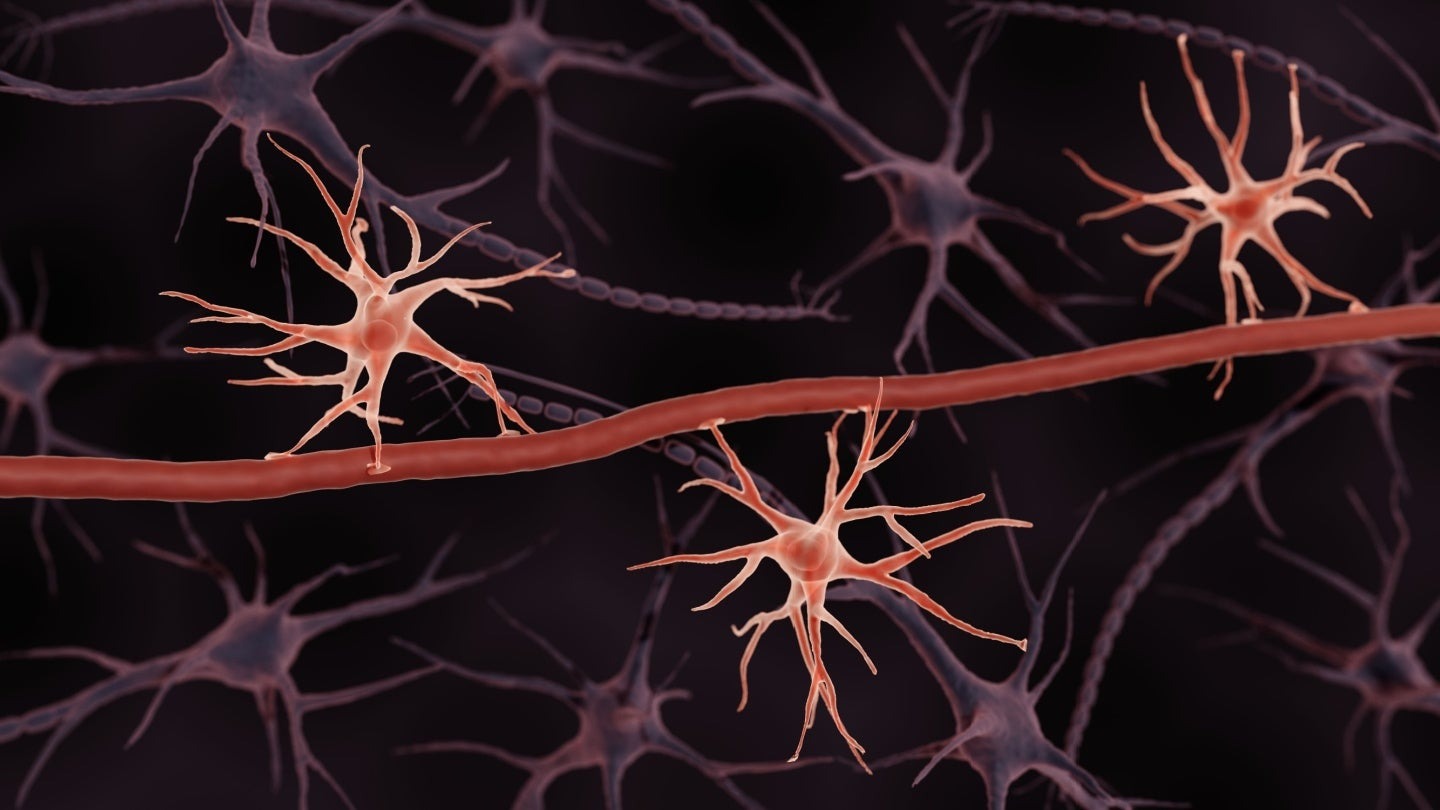
Astrocytes are a type of neural cell that builds the BBB, and Excellio plans to derive exosomes from them to make them even better at targeting the brain. Credit: ART-ur / Shutterstock
Caption. Credit:

Phillip Day. Credit: Scotgold Resources
Total annual production
Australia could be one of the main beneficiaries of this dramatic increase in demand, where private companies and local governments alike are eager to expand the country’s nascent rare earths production. In 2021, Australia produced the fourth-most rare earths in the world. It’s total annual production of 19,958 tonnes remains significantly less than the mammoth 152,407 tonnes produced by China, but a dramatic improvement over the 1,995 tonnes produced domestically in 2011.
The dominance of China in the rare earths space has also encouraged other countries, notably the US, to look further afield for rare earth deposits to diversify their supply of the increasingly vital minerals. With the US eager to ringfence rare earth production within its allies as part of the Inflation Reduction Act, including potentially allowing the Department of Defense to invest in Australian rare earths, there could be an unexpected windfall for Australian rare earths producers.
