Feature
3D printing in surgery: coming to hospitals soon?
3D printing and its associated technologies are helping to evolve surgical procedures in a range of meaningful ways, writes Ross Law.

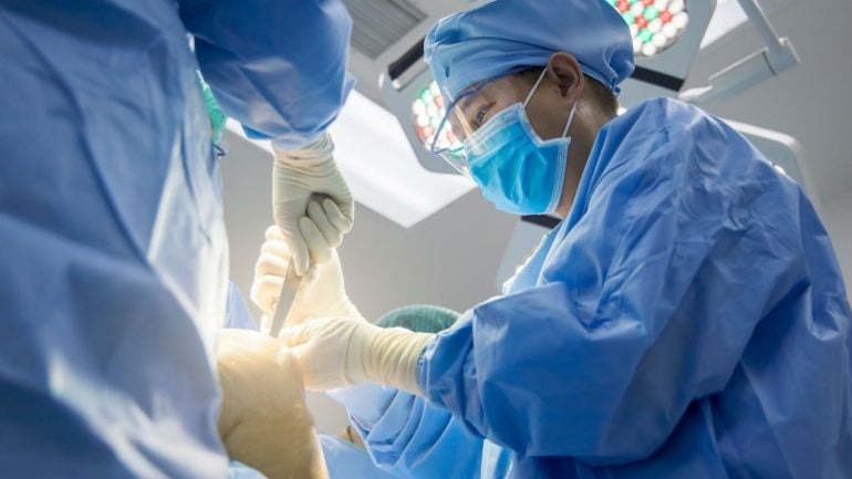
3D-printed surgical guides expedite procedures and increase their accuracy. Credit: Shutterstock / Peter Porrini
3D printing and its associated technologies are gradually taking up a place in the US surgical landscape.
According to GlobalData analysis, factors such as customisation, lower production costs, and quick turnarounds will double the value of the medical 3D printing market from $2bn in sales in 2022 to $4bn in 2026.
One area in which 3D printing is driving change is in the planning of, and assistance during surgical procedures, with past research published in Academic Radiology finding that the use of 3D anatomic models as surgical guides reduced surgical time by a mean of 62 minutes and resulted in cost savings of $3,720 per case.
Another study from 2021 found that surgeries with a duration of four to eight hours were reduced by 1.5 to 2.5 hours if patient-specific instruments were used.
The technology also has applications in the production of medical implants, in which customisability is key.
In-house 3D provisions in hospitals and medical centres across the US are currently not widespread. However, research from 2019 by the American Hospital Association (AHA) found that 113 hospitals in the US had centralised 3D facilities for point-of-care manufacturing, an increase from just three in 2010.
While in-house provisions are gradually rising, there are also moves by 3D printing companies to make 3D-printed equipment more accessible to healthcare providers in the US.
In June 2024, 3D printing company Ricoh launched its flagship Point-of-Care 3D medical device manufacturing facility at Atrium Health Wake Forest Baptist Medical Centre in North Carolina.
The facility aims to provide clinicians with “easy and immediate” access to development, design, and manufacturing services for patient-specific, 3D-printed anatomic models.
Ricoh has also stated that it plans for this facility to be the first of many that are connected to a US healthcare system.
In-house 3D printing comes to Yale
In April 2024, Dr Lisa Lattanza, chair, and Ensign Professor of Orthopaedics & Rehabilitation at Yale School of Medicine (YSM) performed the first fully in-house 3D surgery following the establishment of the 3D Collaborative for Medical Innovation (3DC) in YSM’s Department of Orthopaedics & Rehabilitation.
Having previously relied on external vendors for 3D printing services, with 3DC in-house at YSM, changes can quickly be made to any of its provisions based on real-time feedback from clinicians. They can also work with faster lead times as no shipment of 3D parts is required from third-party vendors.
This first procedure performed by Dr Lattanza was an osteotomy correction for a wrist fracture that healed in a bad position.
Osteotomy is a procedure in which a misaligned bone is cut, moved into a corrected position, and secured in place by a plate as it heals.
For the 3D-printed production of a surgical guide and a plastic model of the patient’s anatomy for reference, the first step is to obtain images of the affected anatomy with a computed tomography (CT) scan.
At YSM, in-house engineer Alyssa Glennon then formats the CT images using software that creates 3D model renderings from stacks of 2D image data. At this stage, the CT image showing the ‘healthy’ side of the patient’s anatomy is mirror-imaged and overlaid on the deformed anatomy to evaluate where the changes are.
Once we see where those changes are, we can plan where we’re going to cut the bone.
Dr Lattanza
“We do this all virtually on the computer, so we can make the osteotomy in multiple different places to decide where the most appropriate place to do the correction is.”
From here, relevant standard orthopaedic plates are selected for use in the surgery.
Following these preliminaries, surgical guides, jigs, and plastic models of the patient’s anatomy, in this first case the radius, are 3D printed and then sterilised for use in surgery.
“We make sure that the guide fits the bone in the patient exactly the way we planned for it to fit on the plastic bone. Once we have made sure that’s the case, we secure the guide to the bone with wires, and then we do whatever the plan has been,” says Lattanza.
In osteotomy, such plans generally involve drilling holes and then making the necessary bone cuts.
The great thing about this approach, Lattanza states, is that all that needs to be done to ensure the correction has been completed as planned during the surgery is to line up those holes.
She explains: “If the bone is rotated off 90° and when we drill those holes, they’re off 90° on the bone, we make the cut then we rotate and line up those holes to put the plate on because the plate holes are straight, and that’s how we know that we’ve got the correction.”
Beyond making relatively common osteotomies more accurate, a 3D provision also allows for more complex cases to be worked upon. Lattanza relays a recent case in which a child had broken the radius and ulna bones in their forearm.
“During the time that she was growing, this deformity got ‘very 3D’, meaning it was off in the sagittal, coronal, and axial plane,” says Lattanza.
“You can’t see the axial plane on an X-ray, and if you can’t see it, you can’t correct it.”
In this case, the procedure required two cuts in the radius to restore it to normal anatomy, and one in the ulna.
“In my career prior to having the 3D technology, that’s something that is difficult or impossible to plan and to execute in the operating room, because you wouldn’t even be able to see that you needed two cuts to make it normal again,” explains Lattanza.
Lattanza is keen to add that the influence of 3D printing on preoperative planning and during surgery should not be a cause for complacency, particularly given that there remain limitations to 3D visualisations of CT scans, chiefly in that the current technology cannot show soft tissue.
“Some people think that this is kind of a phone it in now, but that’s not how it works,” she says.
“This is a collaboration between an engineer and a surgeon, and it has to be that way to get a good result.”
When off-the-shelf implants are not an option
In addition to using 3D-printed surgical guides, Dr Brandon Jonard, orthopaedic oncology specialist at University Hospitals (UH), Cleveland, has used 3D printing for several years to create implants.
While rare, custom 3D-printed implant procedures are undertaken when a patient needs reconstruction in a region of the pelvis, including the hip bones or the hip socket (acetabulum), following the removal of a bone tumour (tumour resection).
“Most hip prostheses that we use to reconstruct somebody’s acetabulum are designed for people with arthritis, but there really isn’t anything developed off-the-shelf to deal with massive bone loss, which is what we end up creating when we do tumour resections,” explains Jonard.
Each of these bone defects is unique, which is why we come into these situations of needing custom implants.
3D-printed surgical guides are of particular value in pelvic osteotomies since they are often complicated in nature and may require multi-planar osteotomies, where cuts are being made on two different planes.
“If you’re going to do a reconstruction on these patients, then the implant that you’re going to utilise has been custom-made to that patient, and it is also custom-made to the osteotomy that you planned,” says Jonard.
“It doesn’t do you any good to make a custom implant and then make the bone cut in someplace different, because then the implant just won’t fit.”
The future of 3D technology in surgery
Looking ahead, Jonard believes implants and customised cutting jigs will become more prevalent in orthopaedic oncology procedures, not only for the pelvic and periacetabular region but also in certain long bone defects and potentially in areas where tissue transplantation (allograft) has been used previously.
He also notes that in orthopaedics as a whole, 3D printing is becoming more widely acceptable for creating complicated implants but also for equipment that is more standard of care.
“Things like acetabular cup components are becoming more commonly 3D printed and one day I imagine that total knee prostheses may become 3D printed,” says Jonard.
We may even find applications where we create customised plates or nails that we use very commonly now, in scenarios where one of these tools off the shelf is unsuitable for use.
While acknowledging that 3D printing surgical guides for osteotomies in-house is a big step, Lattanza believes that future 3D technology used for the visualisation side of surgical planning will integrate with augmented reality (AR) and virtual reality (VR) to undertake these procedures.
“Right now, we’re doing it by printing plastic, but the future of this is to be using AR and VR to do these kinds of osteotomies, getting rid of the printed plastic and using other technology so we can go in the operating room with AR technology, see the deformity in real-time, and make our cuts based on what we’re seeing on a screen,” she says.
Once this is possible, Lattanza foresees the technology providing remote virtual assistance on a global scale, and helping those who don’t have access to the technology.
“It’s a health equity issue, as well as a technology issue. And that’s a really exciting part of it,” she says.
In support of this future, Yale introduced a Masters of personalised medicine and applied engineering programme in 2022. The one-year degree covers 3D preoperative surgical planning technology and related areas that Lattanza views as currently lacking in training in medicine and engineering.
“We want to make sure that we’re on the cutting edge and that we’re prepared for and keeping up with the technology and using it to the best advantage for our patients,” she concludes.
Clinician comfortability and access to 3D printing and related technologies look set to be the factors on which its wider adoption will depend. Based on GlobalData’s projected value of the medical 3D printing market, it seems clear that this is happening in real-time.
The news that YSM has established an in-house provision for 3D printing has turned heads. It may prove to be a turning point in the way people view 3D printing, once they realise what it can be used for, and as access to 3D printing tools and technology continue to improve, with the enactment of healthcare-connected 3D equipment facilities such as Ricoh’s North Carolina site, the wider adoption of 3D printing in the surgical space seems assured.

Astrocytes are a type of neural cell that builds the BBB, and Excellio plans to derive exosomes from them to make them even better at targeting the brain. Credit: ART-ur / Shutterstock
Caption. Credit:

Phillip Day. Credit: Scotgold Resources
Total annual production
Australia could be one of the main beneficiaries of this dramatic increase in demand, where private companies and local governments alike are eager to expand the country’s nascent rare earths production. In 2021, Australia produced the fourth-most rare earths in the world. It’s total annual production of 19,958 tonnes remains significantly less than the mammoth 152,407 tonnes produced by China, but a dramatic improvement over the 1,995 tonnes produced domestically in 2011.
The dominance of China in the rare earths space has also encouraged other countries, notably the US, to look further afield for rare earth deposits to diversify their supply of the increasingly vital minerals. With the US eager to ringfence rare earth production within its allies as part of the Inflation Reduction Act, including potentially allowing the Department of Defense to invest in Australian rare earths, there could be an unexpected windfall for Australian rare earths producers.
